500,000
Annual Readers
1,000
Experts interviewed
200,000
professionals insured
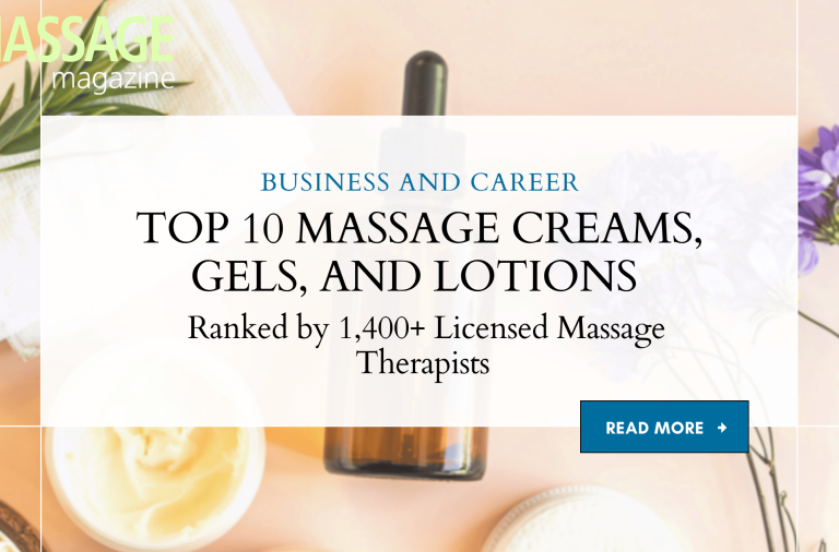
In 2025, MASSAGE Magazine surveyed professionals across private practices, spas, and mobile settings to identify the top-performing massage creams, gels, and lotions in real treatment rooms.
500,000
Annual Readers
1,000
Experts interviewed
200,000
professionals insured
1-YearPart-time Membership
$159
For those who work <10hrs. per week
1-YearFull-time Membership
$169
Our most popular policy option
2-YearMembership
$299
Our most affordable policy option.
Continuing EducationMembership
$59
Massage StudentMembership
$49
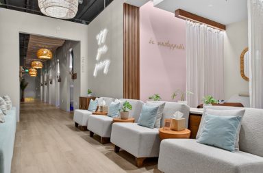
Welcome to our Professional Resource Centers, your one-stop hub for career advancement. Discover a curated suite of resources, tools, and insights designed to boost your professional journey.

The Harmony DX™, an eco-friendly yet economically priced full size table, is crafted from high quality hard Maple from managed forests and finished with earth-friendly, water-based lacquer and glues. Layered with our lightweight yet responsive CFC-Free 2 ½” cushioning system and wrapped in Nature's Touch upholstery adds comfort, durability and style.
Package includes: Harmony massage table, Deluxe Adjustable Headrest and single pocket carry case